Adora
Take a step into the future with Adora and be inspired by the smooth, precise and rapid movements that bring workflow, ergonomics and examination excellence to a completely new level.
Every element and functional aspect of the Adora has been designed with end users in mind. Through a continuous development collaboration between users, engineers and designers, Adora combines outstanding technical quality with a unique approach to user friendly design.
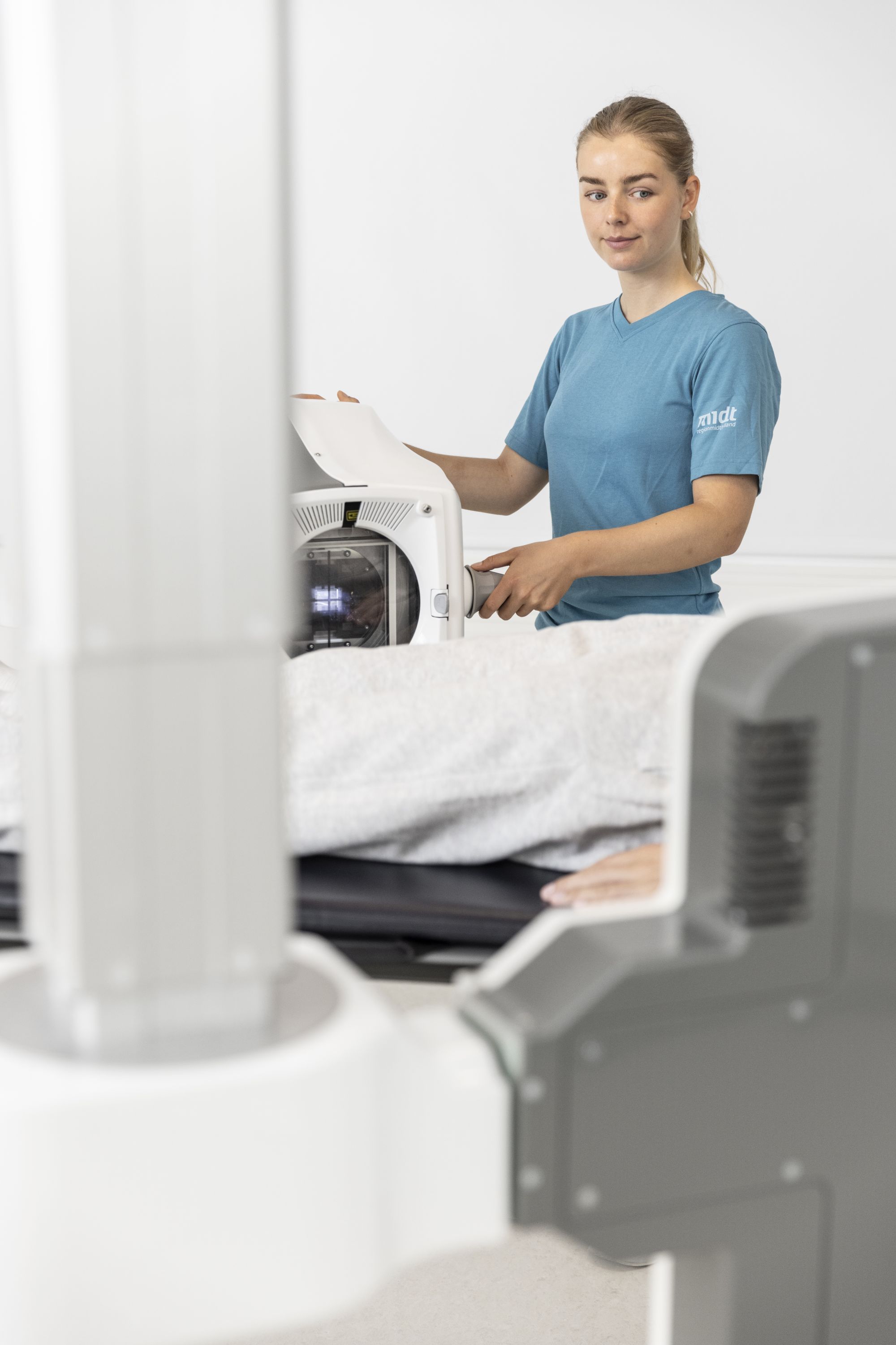
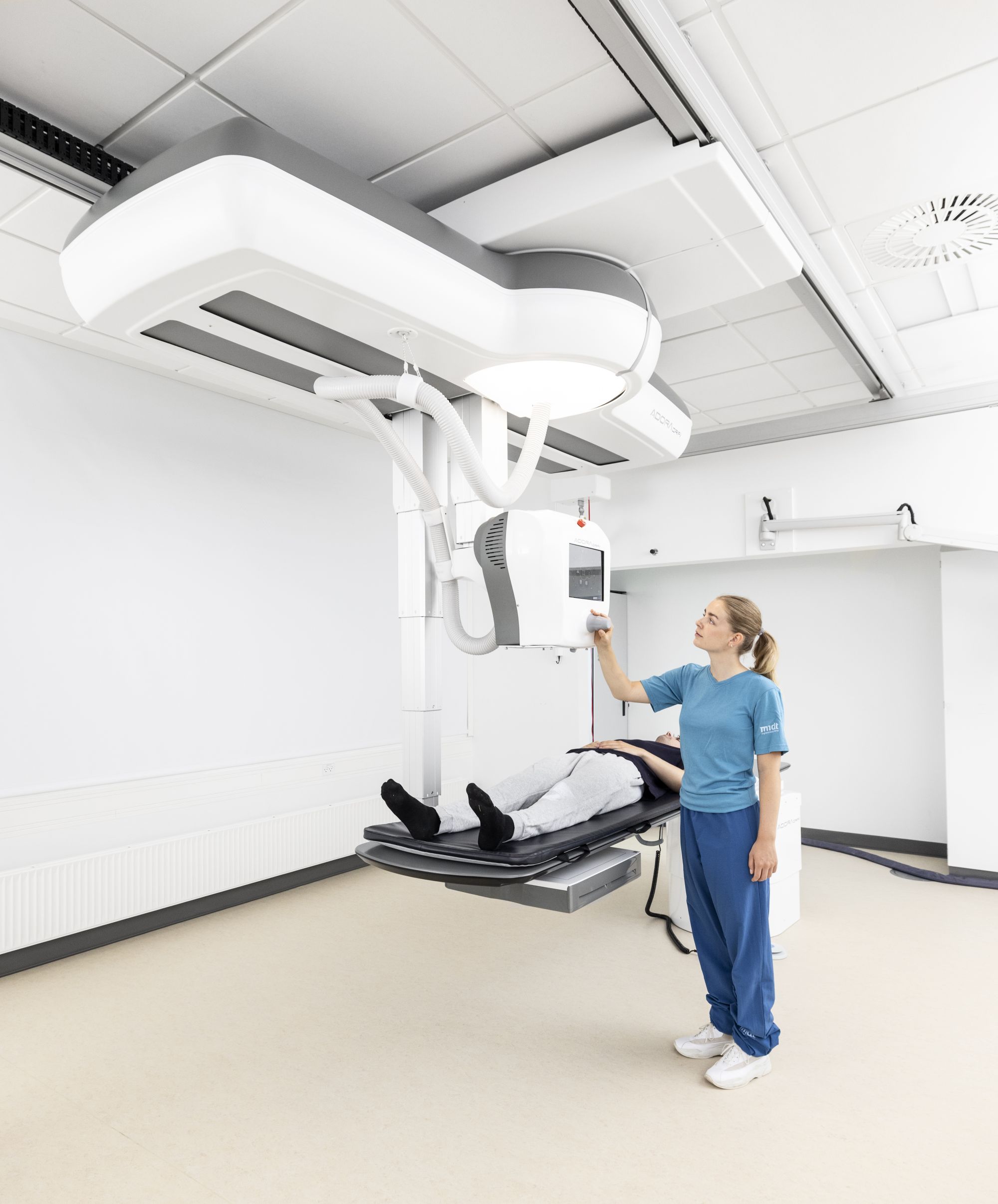
Adora is an automatic, motorized digital radiographic imaging solution featuring a unique, rotating ceiling unit with two telescopic arms, one for the X-ray tube and one for the detector; this allows you to perform exposures from practically any angle through one-touch, stress-free operation.
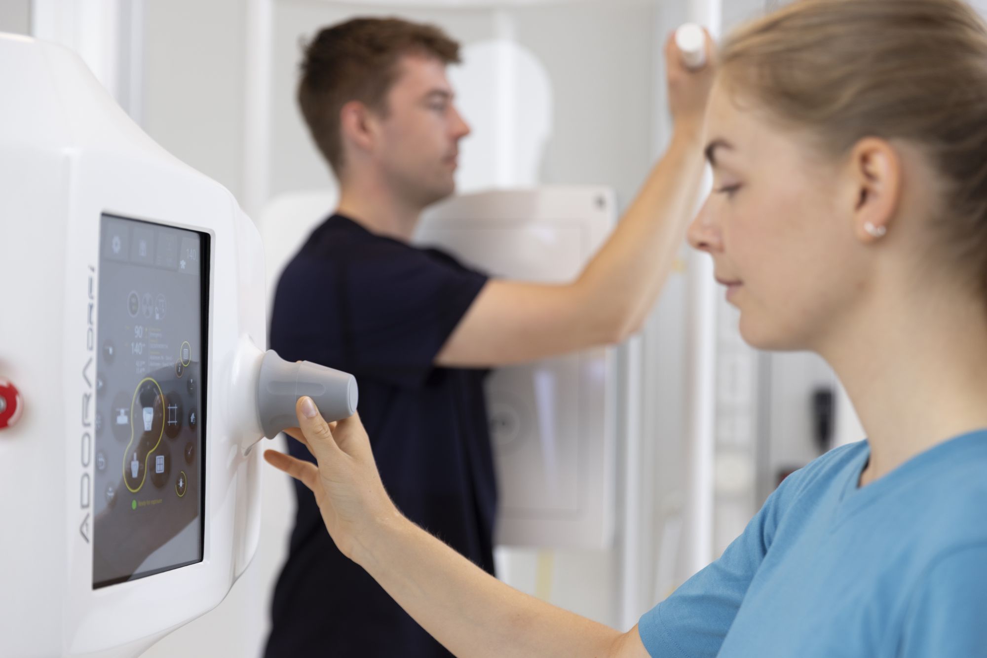
Imagine any projection and perform it: exploit the system’s extensive movement ranges and many detector-to-tube tracking options. The inMotion auto-positioning technology enables fast and intelligent automatic positioning of the detector and tube to any saved position with all examination parameters pre-set.
Inspired by users, the Adora offers the ideal way to improve health care delivery and is available in two different versions: Adora DRi and Adora DRFi.
Save and restore any position – permanently or on-the-fly. Preferred positions can be saved and recalled at any time, for fast and fully automatic positioning.
OUTSTANDING IMAGE QUALITY
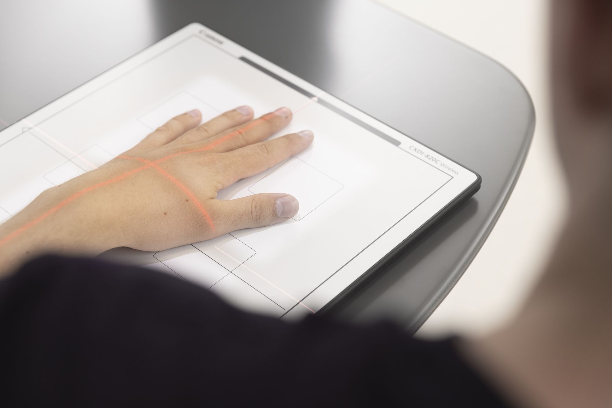
A wide range of high-quality flat panel detectors from Canon is the cornerstone in Adora system configurations. The Canon detectors and CXDI software provide fast, high quality, digital image capture on demand and cover an extensive range of radiographic examinations.
With Adora, one detector is all you need. In addition to the docked detector, Adora supports up to two portable detectors. Thus, ergonomics and examination flexibility can be further enhanced by adding a wireless detector to the configuration.
Intuitive Canon software provides sophisticated image processing for premium diagnostic image quality and can be supplemented with options such as Scatter Correction (reduces dose, eliminates grid artefacts) or Advanced Edge Enhancement (enhanced contrast and detail visibility). The extensive portfolio of Canon wireless detectors ensures you the highest performance in workflow, sensitivity, image quality and versatility, and can even be shared between rooms/modalities.*


Special imaging functionalities
inOrbit
inOrbit makes the tube and detector rotate on an imaginable sphere around either a horizontal or vertical anatomy at an SID specified by the user, and is highly useful in examinations where multiple precision images of the same anatomy are needed.
inTurn (Adora DRFi only)
User configurable, pre-programmed positioning mode by which a 90-degree position change can be executed within 10 seconds. Applicable in connection with e.g., myelographic procedures where the alternate dual perspective is paramount for the clinician.
Scatter correction
Canon’s Scatter Correction option reduces the effect of scattered radiation for non-grid examinations, allowing images with outstanding contrast while avoiding the grid handling and improving the workflow.*
See images here
Advanced Edge Enhancement
Canon’s Advanced Edge Enhancement image processing is designed to enhance the visualization of tubes, catheters and bone details. Besides the original diagnostic image, additional companion views can be added for a specific diagnostic or clinical purpose. Advanced Edge Enhancement filter type and effect can be pre-defined in each protocol*.
See images here
* Product documentation with references is available upon request.
Adora delivers a remarkable level of examination flexibility and capacity utilization.